The microscopes are part of the Technion Center for Electron Microscopy of Soft Matter.
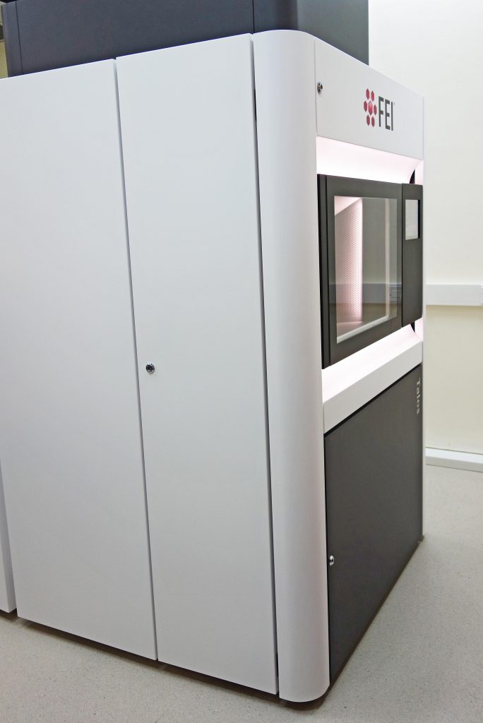
FEI Talos 200C

The FEI (now Thermo Fisher Scientific) Talos 200C, is a cryo-dedicated, high-resolution field-emission gun (FEG)-equipped transmission electron microscope with a demonstrated resolution of 0.12 nm. It allows us to to perform tomography in the conventional TEM and the scanning-transmission electron microscopy (STEM) modes. This state-of-the-art TEM is also equipped with a novel “phase-plate”, which converts phase differences between areas of the specimen to amplitude differences, thus enhancing image contrast without resolution loss, in specimens of inherent little contrast. A Falcon III direct-imaging camera allows imaging at very low electron exposure, essential for electron-sensitive specimens, namely, most of our specimens.
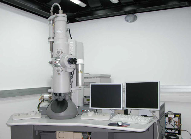
FEI Tecnai T12 G2 Transmission Electron Microscope

This 120 kV, LaB6-emitter equipped, FEI T12 G2 cryo-dedicated transmission electron microscope (installed 2004), is equipped with a Gatan cooled-CCD bottom-mounted, 2048×2048 pixel UltraScan 1000 camera and Gatan 626 cryo-holders and a transfer-station system. It is used as an entry-level TEM and for room-temperature work.
We hope to have this TEM replaced soon.
Zeiss Ultra Plus HR-SEM

This high-resolution scanning electron microscope (HR-SEM) was installed in the Chemical Engineering Sobell Building in February 2008. Its Schottky field emission electron gun provides excellent brightness, even at very low electron acceleration voltages, down to a few tens of volts. This is an important feature for high-resolution imaging of surface nanostructures, and for overcoming charging of uncoated non-conductive specimens. The microscope is equipped with four imaging backscattered and secondary electron detectors: two of the in-lens type, and two outside the column. It is also equipped with a Bruker Xflash x-ray energy dispersive spectrometer (EDS) for x-ray elemental microanalysis. A Leica cryo-stage (installed in 2022, replacing the original Bal-Tec one) allows imaging of cryogenic specimens, such as fast-cooled liquids and biological systems. All this makes a versatile and flexible tool for the complete nano- and microstructural analysis of a wide range of systems.
By the end of 2022, we expect to install a new out-of-the-column backscattered electron detector (AsB detector in the Zeiss lingo), which will allow operation below an acceleration voltage of 1 kV and a room-temperature specimen airlock made possible by the above-mentioned replacement of the cryo-stage.
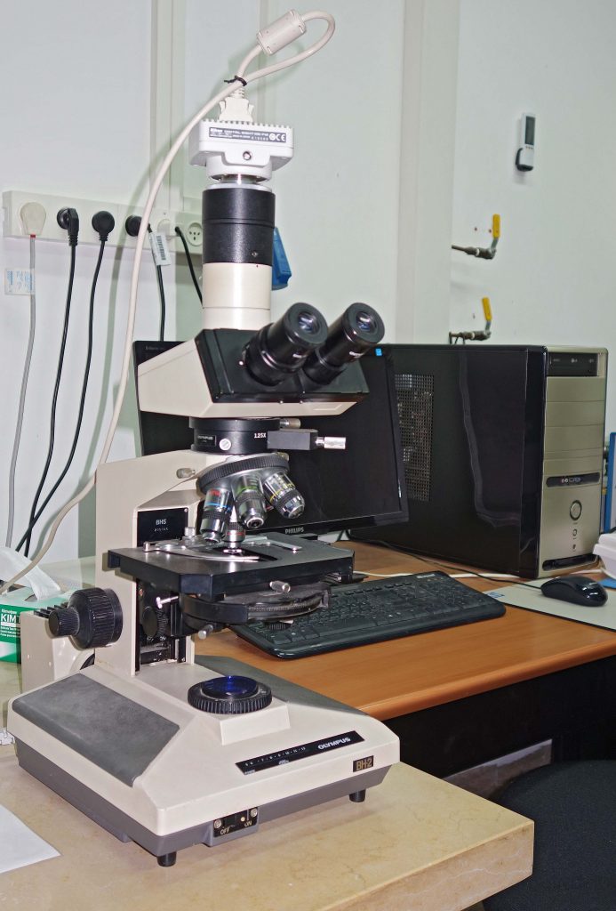
Olympus BH2 light microscope, connected to a Nikon DS-F12 CCD camera

The microscope is equipped for differential interference contrast (Nomarski) optics, phase-contrast, and polarized-light microscopy.
